AMRC
Advanced Material Research Center
INDIAN INSTITUTE OF TECHNOLOGY MANDI
- 01905-267027
- amrcoffice@iitmandi.ac.in
About Instrument and its applications
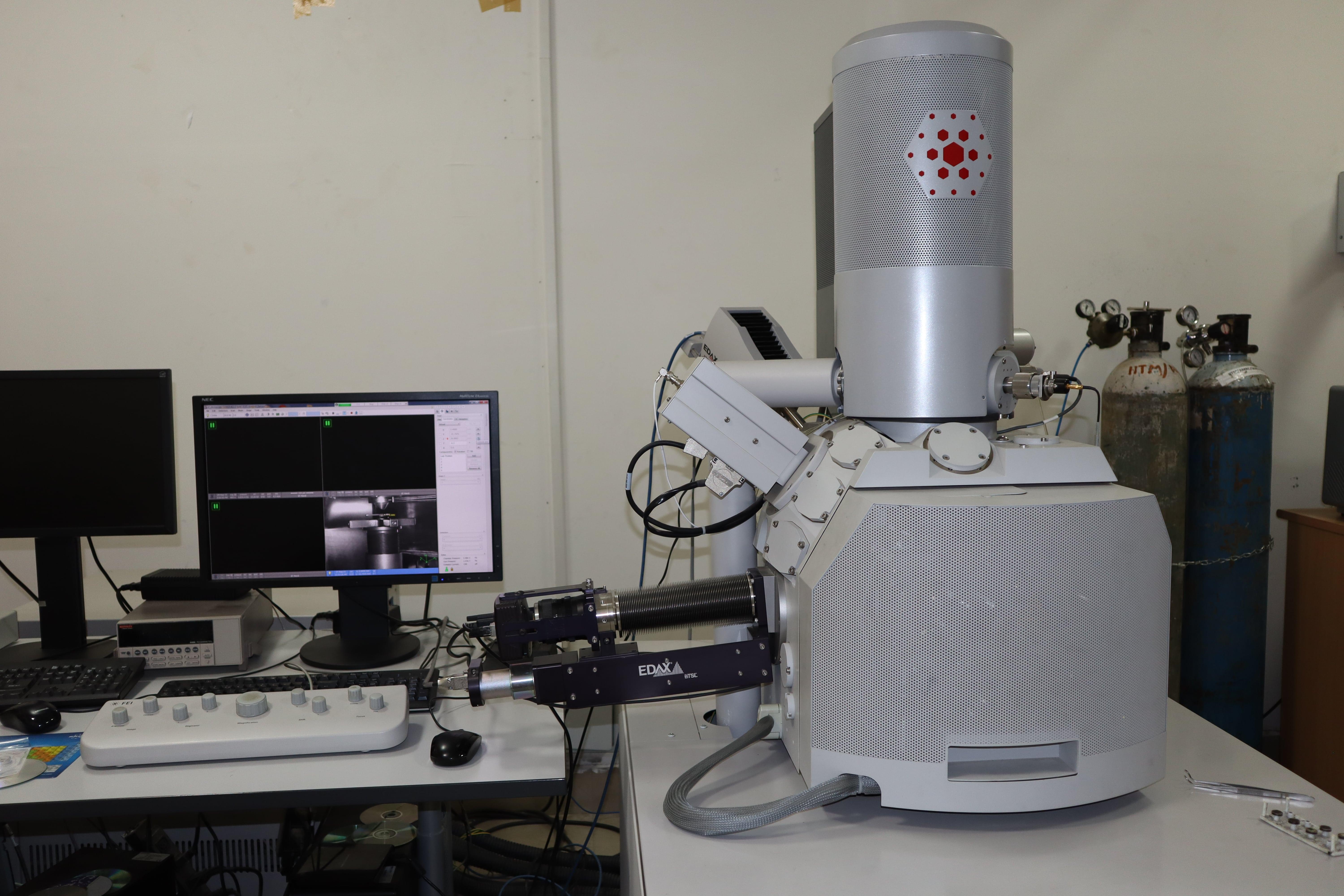
The NanoSEM 450 is a field-emission scanning electron microscope (FE-SEM), that attains ultra-high imaging resolution without the specimen size restrictions of a conventional in-lens FE-SEM due to the advanced design of the electron optics. The NanoSEM 450’s Schottky field-emission source allows the user to achieve high imaging resolution at a range of kV, at both low (high-resolution imaging) and high (microanalytical imaging) currents. Secondary electron (SE) imaging can be undertaken in both field-free and immersion mode for comprehensive low-to-high-resolution imaging of a variety of samples. The NanoSEM 450 is fitted with a retractable annular backscattered electron detector as well as an EDAX SDD-EDS detector for the convenient visualization of compositional differences across the specimen surface.
General Information
Make- FEI(currently supported by Thermo Fisher)
Model- Nova Nano SEM 450
Year of installation- 2013
Sample Requirements
Powder/Solid samples
Dry and vacuum compatible sample
Specifications
1. Accelerating voltage – 500v-30 kV
2. Electron Source- FEG with ultra-high brightness Schottky field emitter
3. Resolution- 1.5 nm
4. Magnification range- 30X-100KX
5. Detectors-EDS, EBSD, BSD, LVD