AMRC
Advanced Material Research Center
INDIAN INSTITUTE OF TECHNOLOGY MANDI
- 01905-267027
- amrcoffice@iitmandi.ac.in
About Instrument and its applications
This surface analysis UHV system is fully configured for operation using dedicated Electron Spectroscopy for Chemical Analysis. The analysis chamber is equipped with a full 180° hemispherical, mu-metal shielded analyzer and a 128-channel, signature-corrected, position-sensitive detector.
• X-ray source XPS (X-ray photoelectron Spectroscopy) can determine the elemental composition, empirical formula, chemical state, and electronic state of the elements within a material. XPS spectra are obtained by irradiating a solid surface with a beam of X-rays while simultaneously measuring the kinetic energy of electrons that are emitted from the top 1–10 nm of the material being analyzed.
• UV Source UPS (Ultraviolet Photoelectron Spectroscopy) is used for measuring Valence Band and Work Function.
• REELS (Reflected Electron Energy Loss Spectroscopy) are used for band gap measurements.
• Monatomic and Gas Cluster Ion Source (MAGCIS) are used for sample surface cleaning.
• ISS (Ion Scattering Spectroscopy): It is a technique in which a beam of ions is scattered by a surface and the kinetic energy of the scattered ions is measured.
• Angle-Resolved XPS (AR-XPS):- The information depth for XPS is a few nanometers, depending upon the kinetic energy of the electrons and the material being analyzed. Angle-resolved XPS (ARXPS), however, is a technique that varies the emission angle at which the electrons are collected, thereby enabling electron detection from different depths. ARXPS provides information about the thickness and composition of ultra-thin films.
• XPS Imaging and Mapping:- It is used to identify points or small features at the surface.
• Flood Gun:- When X-ray photons strike a surface, they cause the emission of electrons. This, of course, is the basis of the XPS technique. If the surface is electrically insulating then the emission of electrons causes a positive charge to accumulate at the surface. For this reason, it is necessary to neutralize the charge on the surface by replenishing electrons from an external source. This stabilizes and controls the charging to within a few electron volts of the neutral state.
Facilities:-
• X-Ray Photoelectron Spectroscopy Analysis (XPS)
• Large area XPS Analysis (400 µm Spot Size)
• Micro-area XPS Analysis (10 µm Spot Size)
• XPS Depth Profiling Analysis
• UV Photoelectron Spectroscopy Analysis (UPS)
• Monatomic and Gas Cluster Ion Source (MAGCIS)
• XPS Imaging and Mapping
• Angle-Resolved XPS (AR-XPS)
• Ion Scattering Spectroscopy(ISS)
• Flood Gun ( for Charge Compensation)
• Reflected Electron Energy Loss Spectroscopy (REELS)
Samples Requirements:-
• Size of Thin-film Samples:- 20 mm X 20 mm.
• Size of Pellet Samples:- 7mm to 10mm.
• Thickness of Pellet/Film Samples:- 5mm to 10mm.
• Amount of Powder Samples:- 5mg to 20mg.
General Information
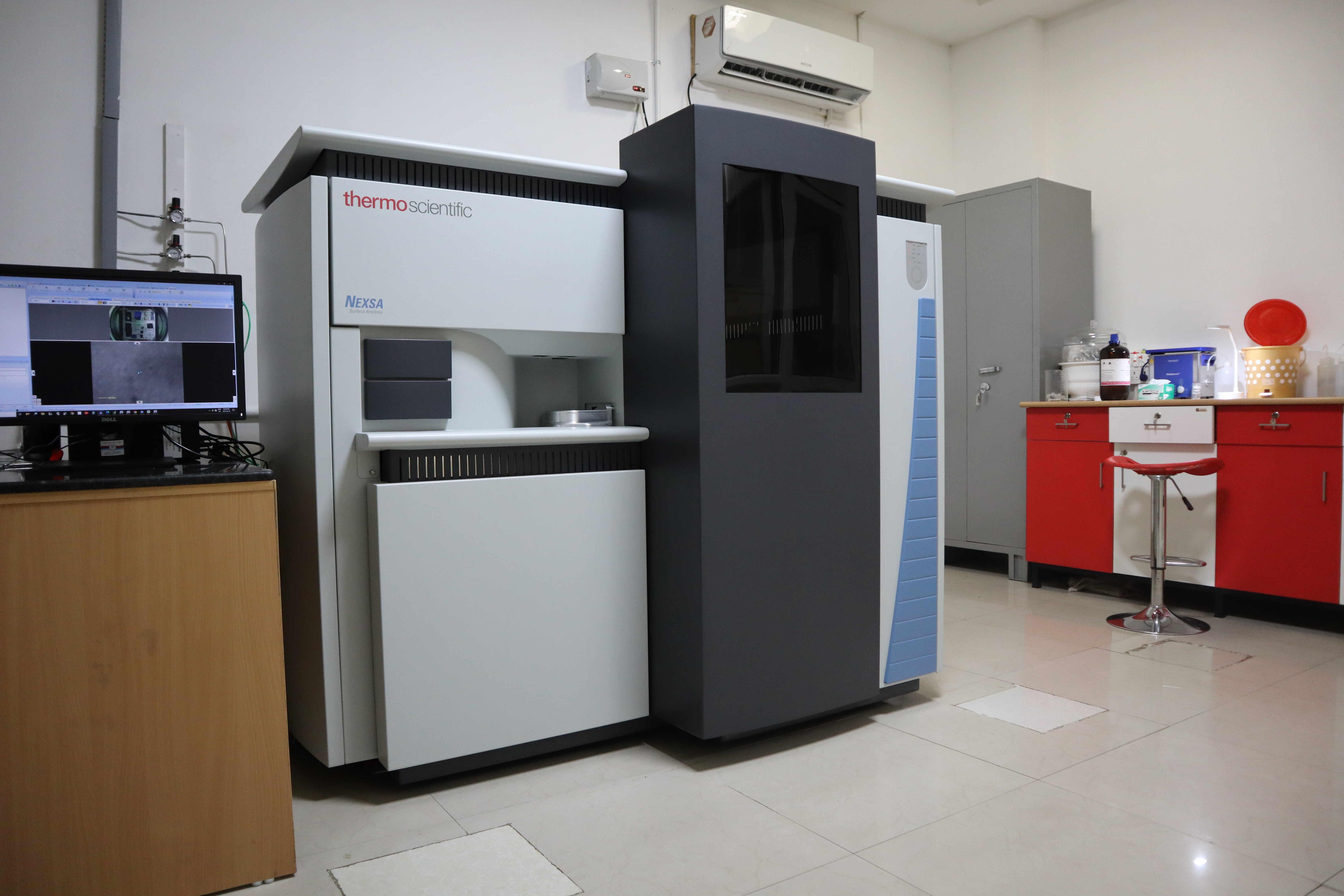
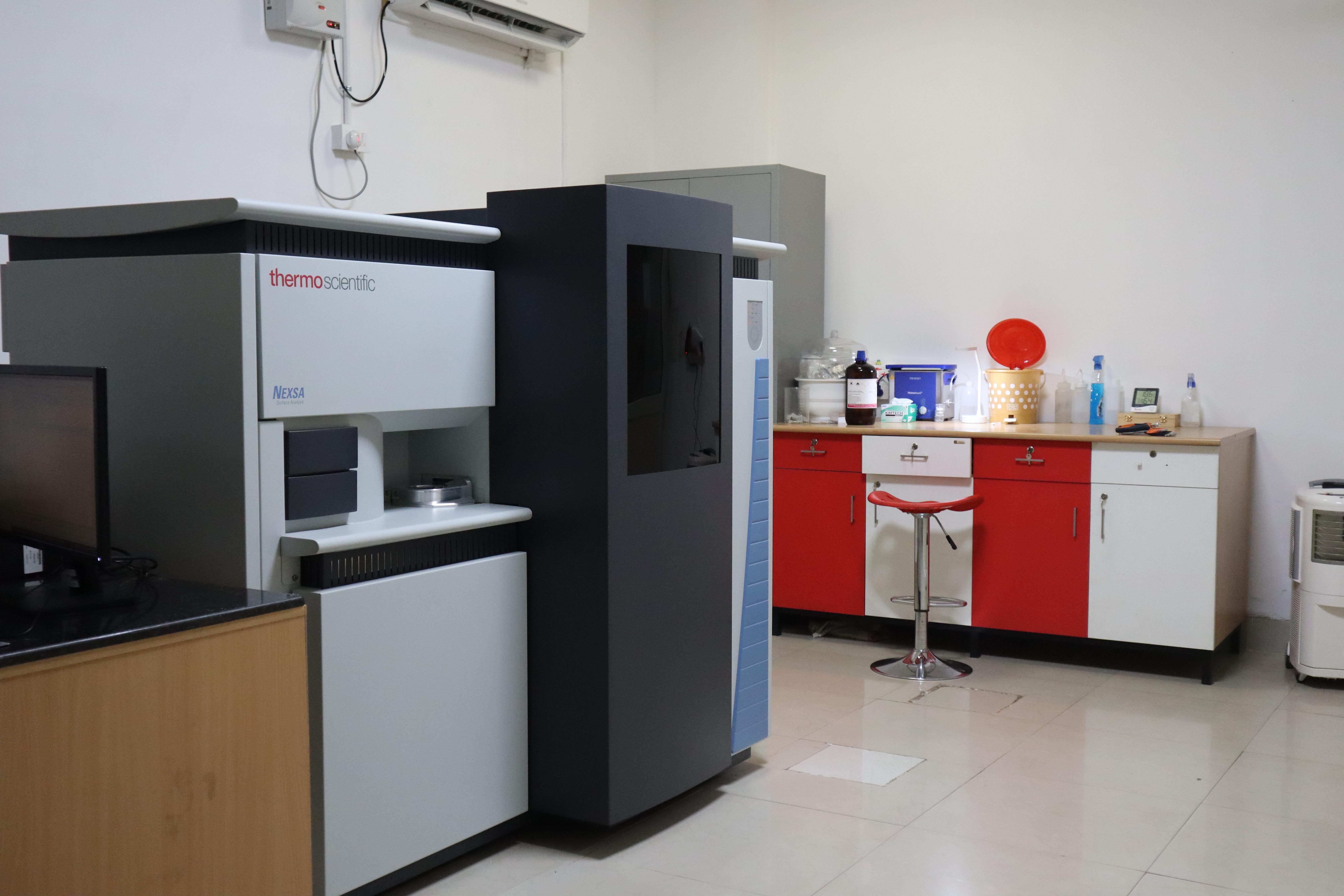
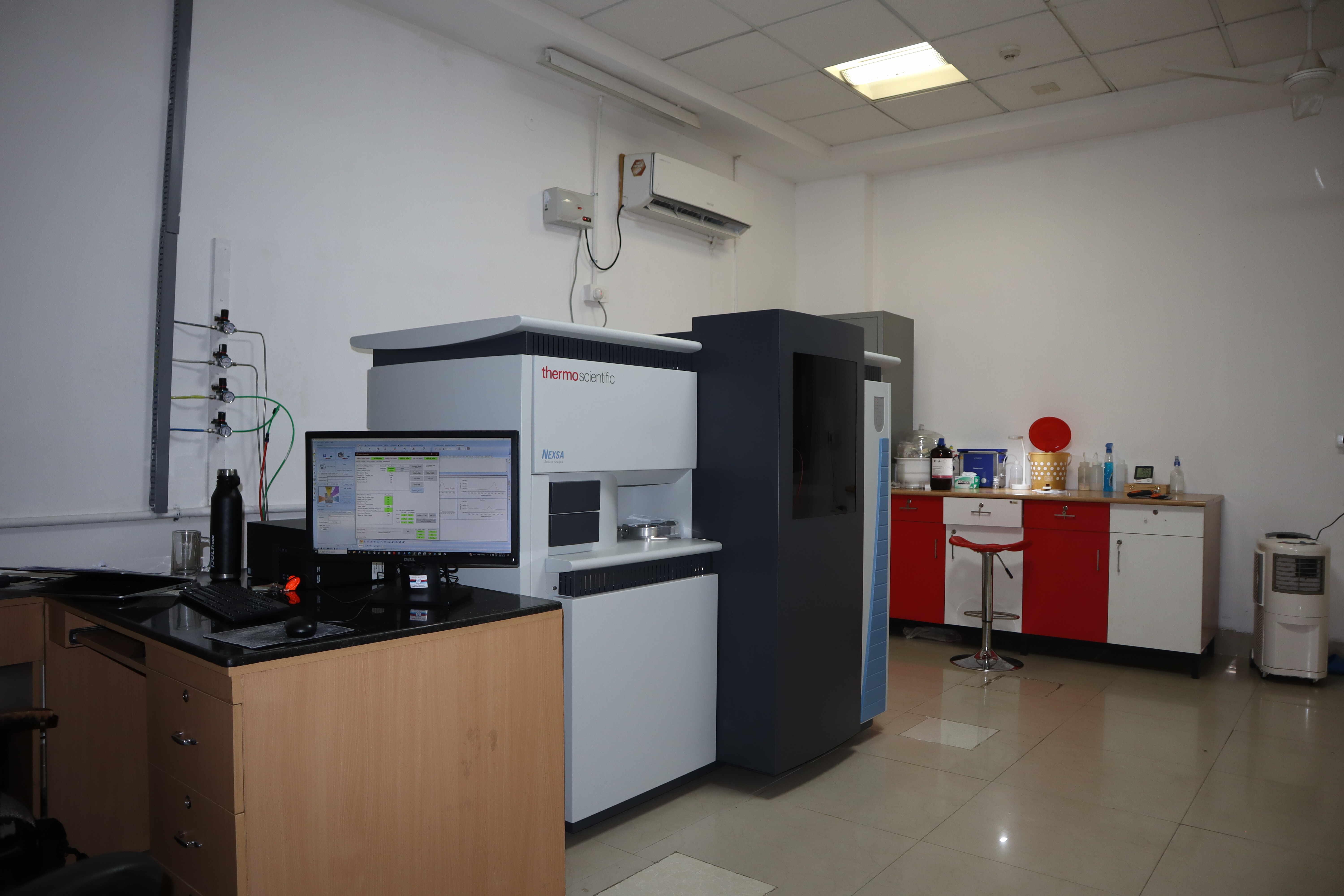
Make- Thermo Scientific
Model- NEXSA Surface Analysis
Specification:-
1. X-Ray Source:- Micro-focused, monochromated Al K-alpha (1486.6eV) X-ray source.
Adjustable x-ray spot size – Maximum spot size 400 µm – Minimum spot size 10 µm.
2. UV Source:- HeI (21.2eV) HeII (40.8eV)
3. REELS Source:- Beam Energy (1000 eV)
4. MAGCIS Ion Source:-
• Ion Energy for Monatomic Ion Source:- 300V to 4000V
• Ion Energy for Gas Cluster Ion Source:- 2000V to 8000V
• Cluster for Gas Cluster Ion Source:- 75 to 2000 atoms
Gas Cluster Use for Polymer Samples
5. ISS Source:- Ion Energy (1000V)
System Base Pressure:-
• Load Lock Chamber: 5.0 X 10-08mbar.
• Analysis Chamber: 3.0 X10⁻¹⁰mbar.